Skip to content

Chapter 31

Biomedical signals

Two lessons in Part XI, Applications. Read them in order, or start anywhere: a prerequisite is a link, never a gate.

Start with 31.1
0 of 2 readabout 34 minutes

Lessons in this chapter

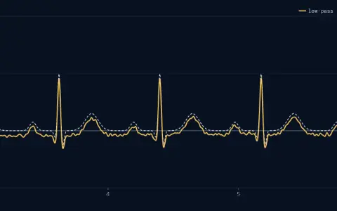
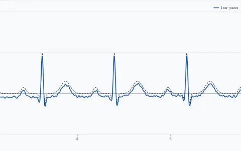
One panel, millivolts from −1 to 2 against time in seconds from 3 to 6. The clean ECG is dashed and labelled clean, with R spikes at 3.62, 4.40, 5.17 and 5.95 s. The solid line, labelled low-pass, is the trace after the high-pass, the notch and the low-pass, from −0.31 to 0.93 mV. Its error RMS against the clean ECG over seconds 1 to 9 is 82 µV.One panel, millivolts from −1 to 2 against time in seconds from 3 to 6. The clean ECG is dashed and labelled clean, with R spikes at 3.62, 4.40, 5.17 and 5.95 s. The solid line, labelled low-pass, is the trace after the high-pass, the notch and the low-pass, from −0.31 to 0.93 mV. Its error RMS against the clean ECG over seconds 1 to 9 is 82 µV.

Lesson 118 minYou are hereRead

ECG processing

Clean a synthetic ECG with a high-pass, a notch and a low-pass, then find its beats with Pan–Tompkins and read the heart rate.

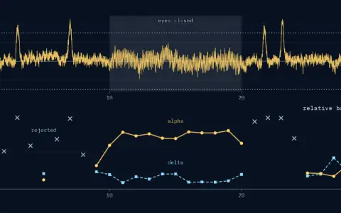
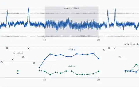
Two stacked panels over 0 to 30 s. EEG panel: the synthetic record in µV, from −46.3 to 111.5, against dotted reject levels at plus and minus 75 µV; four blinks rise above the upper one; the eyes-closed span, 10 to 20 s, is shaded. Relative band power panel: 29 windows of 29 drawn, centred at 1 to 29 s; alpha's share as dots on a solid line, delta's as squares on a dashed line; 10 rejected windows (centred at 2, 3, 4, 6, 7, 8, 21, 22, 23 and 24 s) shown as open crosses at their delta share. Span 20 to 30 s: alpha 22 %, delta 28 %, mean over the kept windows.Two stacked panels over 0 to 30 s. EEG panel: the synthetic record in µV, from −46.3 to 111.5, against dotted reject levels at plus and minus 75 µV; four blinks rise above the upper one; the eyes-closed span, 10 to 20 s, is shaded. Relative band power panel: 29 windows of 29 drawn, centred at 1 to 29 s; alpha's share as dots on a solid line, delta's as squares on a dashed line; 10 rejected windows (centred at 2, 3, 4, 6, 7, 8, 21, 22, 23 and 24 s) shown as open crosses at their delta share. Span 20 to 30 s: alpha 22 %, delta 28 %, mean over the kept windows.

Lesson 216 minYou are hereRead

EEG and rhythms

Read a synthetic EEG by frequency band: alpha rises as the eyes close, blink windows are rejected, and averaging trials recovers a small evoked response.

After this chapter

Where to go next.

The chapters either side, and the rest of Part XI in the library.

Phasorium
LibraryEvery lesson, in order

Parts

About Phasorium
Look